
Introduction
Pregnancy brings many questions about whether a baby is growing normally and developing as expected. Ultrasound is an important part of prenatal care because it allows healthcare professionals to assess the pregnancy without using radiation. When more detailed imaging is appropriate, 3D and 4D ultrasound can provide additional views of the baby and certain anatomical structures.
For patients looking for the best 3D-4D sonography center in Ahmedabad, it is important to understand that 3D-4D sonography is not simply about getting clearer pictures of the baby. Its usefulness depends on the stage of pregnancy, the reason for the examination, the baby’s position, and the quality of the images obtained.
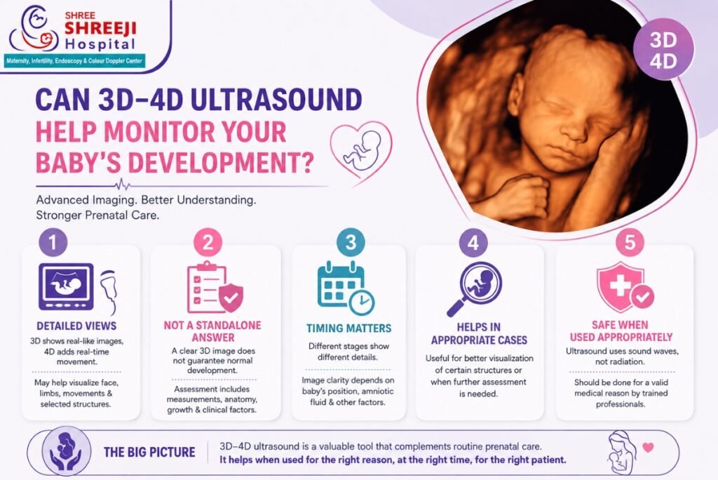
A 3D ultrasound creates a three-dimensional image, while 4D ultrasound adds movement in real time. These technologies can complement routine pregnancy scans when used for an appropriate clinical purpose.
What Can 3D-4D Imaging Show During Pregnancy?
Traditional ultrasound produces two-dimensional images that healthcare professionals use to evaluate fetal structures, growth, and the pregnancy environment. 3D ultrasound processes multiple image planes to create a three-dimensional representation.
4D ultrasound takes this a step further by displaying those three-dimensional images as moving images.
Depending on the pregnancy stage and imaging conditions, these techniques may provide additional visualization of features such as:
- The baby’s facial structures
- Limbs and body contours
- Certain external anatomical features
- Fetal movements
- Selected anatomical structures that may benefit from additional visualization
However, a 3D or 4D image does not automatically provide a more complete assessment of every aspect of fetal health. Standard two-dimensional ultrasound remains an important part of prenatal imaging, and specialized examinations may be required when a particular concern needs closer assessment.
Does Seeing the Baby in 3D Mean Development Is Normal?
Not necessarily.
A detailed-looking image can be reassuring for parents, but the appearance of a 3D picture alone cannot confirm that every aspect of fetal development is normal. Assessment of fetal growth and anatomy involves measurements, imaging from different views, pregnancy history, gestational age, and other clinical information.
A healthcare professional may assess factors such as:
- Fetal growth and measurements
- Development of major anatomical structures
- Fetal movement
- Position of the baby
- Placental location
- Amniotic fluid
- Other findings relevant to the stage of pregnancy
This is why 3D-4D sonography should be viewed as an imaging tool rather than a standalone test that can guarantee a healthy pregnancy.
When Can 3D-4D Ultrasound Add Useful Information?
The value of 3D-4D imaging varies from one pregnancy to another. In some situations, a healthcare professional may consider additional imaging when certain structures need better visualization or when a finding on a routine scan requires further assessment.
It may also help provide a different perspective on selected fetal anatomical features. In some cases, three-dimensional reconstruction can assist the clinician in understanding the spatial relationship of structures.
The decision to use this technology should therefore be based on the medical purpose of the scan rather than simply choosing the most advanced-looking imaging option.
Parents should also understand that image quality can vary considerably. The baby’s position, movement, gestational age, amount of amniotic fluid, and other factors can affect how clearly particular structures are seen.
Why Does the Timing of the Scan Matter?
There is no single week that produces the same type of useful information for every pregnancy.
Different stages of pregnancy allow healthcare professionals to evaluate different aspects of fetal development. Earlier scans may focus on specific developmental milestones, while later examinations can provide information about growth, anatomy, position, and other pregnancy-related findings.
For 3D-4D imaging, the baby’s position can be particularly important. If the baby’s face is turned away, or if another structure is between the ultrasound probe and the area being examined, obtaining a clear image may be difficult.
This means that an unclear 3D image does not necessarily indicate a problem with the baby. It may simply reflect the baby’s position or the conditions during the examination.
Can 3D-4D Sonography Detect Developmental Problems?
Ultrasound can help healthcare professionals evaluate many aspects of fetal anatomy and development, and 3D imaging can sometimes provide additional visualization of certain structures.
However, no ultrasound technique can identify every possible fetal or pregnancy-related condition. Some findings may be difficult to detect because of fetal position, gestational age, image quality, or the nature of the condition itself.
If a routine scan identifies something that requires closer evaluation, your doctor may recommend a targeted or detailed ultrasound examination. Depending on the finding and the pregnancy, additional testing or specialist assessment may also be considered.
The purpose is not to create unnecessary concern but to make sure that any significant finding is evaluated appropriately.
What Should Parents Expect During a 3D-4D Scan?
A 3D-4D ultrasound is generally performed using an ultrasound transducer placed over the abdomen with ultrasound gel. The healthcare professional moves the probe across the area being examined to obtain the required images.
The scan may take varying amounts of time depending on the purpose of the examination and how easily the relevant structures can be visualized.
Parents may see recognizable images of the baby’s face, hands, feet, or movements when conditions are favorable. However, the ability to obtain such images should not be considered the primary measure of the quality or medical value of the examination.
If the baby’s position makes imaging difficult, the healthcare professional may need to obtain views from different angles or continue the assessment according to the clinical purpose of the scan.
Is 3D-4D Ultrasound Safe During Pregnancy?
Ultrasound uses sound waves rather than ionizing radiation. Diagnostic ultrasound has been used extensively in pregnancy, but it should still be performed for appropriate medical purposes by trained professionals using suitable equipment and settings.
The fact that a technology is considered useful does not mean that more scanning is always better. Unnecessary or prolonged exposure should be avoided, particularly when there is no medical reason for additional imaging.
For this reason, patients should follow the recommendations of their obstetrician or qualified ultrasound professional regarding when an examination is appropriate.
If you are considering the best 3D-4D sonography center in Ahmedabad, it is worth focusing not only on the type of machine available but also on whether the scan is being performed for an appropriate clinical reason and interpreted in the context of your pregnancy.
How Does 3D-4D Imaging Fit Into Regular Prenatal Care?
3D-4D ultrasound should generally be understood as part of a broader prenatal assessment rather than a replacement for routine pregnancy care.
Your healthcare professional may recommend different ultrasound examinations at different stages depending on your pregnancy history, symptoms, gestational age, previous findings, and individual risk factors.
A scan can provide valuable information, but the results need to be interpreted alongside clinical findings and other relevant prenatal assessments.
For example, if fetal growth is a concern, measurements and growth trends may be more important than obtaining a detailed facial image. Similarly, if a particular anatomical structure requires assessment, the examination should be planned around obtaining appropriate diagnostic views.
Choosing Imaging Based on Your Pregnancy Needs
When deciding where to have pregnancy imaging performed, patients may naturally focus on image quality and technology. Those factors matter, but they are only part of the picture.
A useful prenatal imaging service should prioritize appropriate examination protocols, careful assessment, clear communication, and accurate interpretation of findings.
Dr. Bhaumik Shah emphasizes individualized patient care, and pregnancy imaging should similarly be approached according to the mother’s and baby’s specific clinical circumstances rather than using the same approach for every pregnancy.
If you are considering the best 3D-4D sonography center in Ahmedabad, discuss with your healthcare professional why the scan has been recommended, what it is intended to assess, and whether additional imaging is medically necessary.
3D-4D ultrasound can offer valuable additional views of a developing baby in selected circumstances. Its greatest value comes when the technology is used appropriately as part of comprehensive prenatal care—not simply because it produces more detailed or memorable images.